CASE DISCUSSION ON 63 YEAR OLD MAN WITH CONSOLIDATION OF LEFT LUNG
This is an online E log book to discuss our patient's de-identified health data shared after taking his/her/guardian's signed informed consent.
Here we discuss our individual patient's problems through a series of inputs from an available global online community of experts with an aim to solve those patient's clinical problems with collective,current,best evidence based inputs.
This e-log book also reflects my patient centered online learning portfolio and your valuable inputs in the comment box is welcome.
Neha Tipparaju
9th Semester, Roll no. 100
Case of 63 year old man with Consolidation of Left Lung
I've been given this case, in an attempt to understand the topic of "patient clinical data analysis" and to develop my competency in reading and comprehending clinical data including history, clinical findings, investigations as well as to come up with a diagnosis and treatment plan.
All the information was obtained from the patient's relative, under the guidance of Dr. Rakesh Biswas sir.
My view of the case:
Chief Complaints:
A 63 year old man, resident of Nalgonda and a labourer by occupation presented with:
Fever since 21 days
Cold and cough since 21 days
History of presenting illness:
Patient was apparently assymptomatic 21 days back. He had taken his 2nd dose of COVID vaccination then and claims that the fever has started since then.
The fever was incidious in onset, high grade and present throughout the day. It did not relieve on medication.
It was not a/w chills and rigour, headache, burning micturition, vomitings or diarrhoea.
The patient also has complaints of cold and cough since 21 days.
The cough is productive, associated with a moderate amount of sputum production. The sputum was mucoid in consistency and yellowish white in colour. It did not have any odour and was not blood stained.
The cough is associated with diffuse chest pain of dragging type.
The patient is not complaining of any shortness of breath, loss of weight or loss of appetite.
Past history:
There were no similar complaints in the past.
The patient is not a k/c/o Diabetes mellitus, Hypertension, Tuberculosis, Asthma or any heart disease.
H/o Chest trauma 30 years back--- Rib fracture was operated and repaired.
No drug history.
Personal history:
Diet: Mixed
Appetite: Normal
Bowel and bladder: Regular
Sleep: Reduced due to pain
No known allergies
Addictions: Alcohol intake since 40 years.
180 mL/day ( 90 mL in the morning and 90 mL in the evening)
Smokes Chutka since 40 years--- 3 per day.
Family history:
No similar complaints
General Examination:
The patient is examined in the sitting position, in a well lit room after taking informed consent.
He is conscious, coherent, cooperative, well oriented to time, place and person.
Thinly built and nourished. On examination of signs:
Pallor: Present. Palmar creases are not seen
No icterus
No cyanosis
No clubbing
No koilonychia
No lymphadenopathy
No generalized edema
No pedal edema was observed.
JVP was not seen to be raised.
Vitals:
BP: 130/70 mm of Hg
PR: 80 bpm
Respiratory Rate: 21 cpm (high)
Tempature: 98.6°F
GRBS: 217 mg/dL (elevated)
SPO2: 99%
Systemic Examination:
Respiratory system:
On Inspection: Chest is symmetrical. Movements appear to be decreased on left side.
Trachea is central in position. No engorged veins scars or sinuses.
On palpation: No tenderness or rise in temperature. Trachea is central in position. Reduced movement on left side. Tactile vocal fremitus is increased in left supra mammary area.
On percussion: Dull note in left upper lobe
On Auscultation: Bilateral air entry is present. No crepitations heard.
CVS: S1 and S2 heart sounds heard. No murmers
Per Abdomen: Scaphoid in shape. Liver and Spleen are palpable.
No guarding
No rigidity
No rebound tenderness
No shifting dullness
Bowel Sounds heard
CNS: Intact
Investigations:
Complete blood picture shows reduced hemoglobin

He was detected as de-novo Diabetes based on random, fasting, post prandialand glycated hemoglobin levels


Liver function tests were also performed.

HR CT scan:


Impression:
Based on this history and investigations, a provisional diagnosis of:
Left sided lung cavity and Consolidation secondary to infection under investigation with Denovo diabetes.
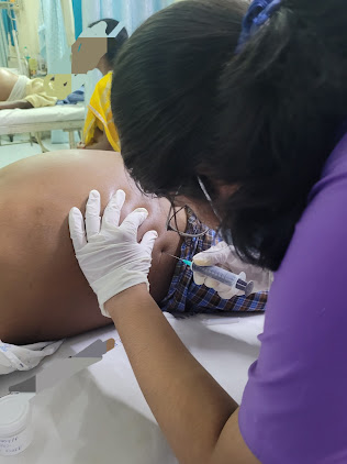
Management:
Patient was put on antibiotics and supportive care

Patient recovered and was discharged.
Comments
Post a Comment